Brachycephalic Ocular Syndrome
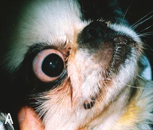
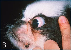
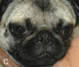
The so called brachycephalic ocular condition (Figure 1), similar to brachycephalic respiratory condition, can be considered a syndrome that—in addition to macroblepharon and lagophthalmos— may include a variety of other factors or clinical signs.1-5 Brachycephalic breeds often have:
- Medial canthal entropion in which the lower eyelid, adjacent to the medial canthus, rolls inward and covers up, or occludes, the lower nasolacrimal puncta2,5,6
- Epiphora and tear staining due to inappropriate tear fluid drainage that, sometimes, are significant enough to cause moist dermatitis ventral to the eye
- Trichiasis or hairs that deviate toward the cornea from the medial caruncle or nasal skin folds adjacent to (sometimes contacting) the cornea;6 this hair chronically irritates the ocular surface—even if the patient does not demonstrate discomfort—and can contribute to keratitis and ocular melanosis.2,3,7 The medial caruncle is a raised nodule of conjunctiva overlying glandular tissue that sits in the corner of the eye adjacent to the medial canthus
- Reduced corneal sensitivity compared to mesocephalic and dolichocephalic breeds;2-4 thus, while afflicted dogs may not exhibit acute or severe corneal pain, diminished innervation can contribute to delayed recognition of the problem and ongoing damage that leads to impaired corneal wound healing
- Quantitative or qualitative tear deficiencies which can further exacerbate corneal injury
- Shallow orbits which offer considerably less protection of the globe than the deeper orbits of their mesocephalic or dolichocephalic counterparts.2-4 This orbital anatomy, especially when combined with excessively long palpebral fissures, greatly increases potential for traumatic proptosis, a globe-threatening condition in which the eyelid margins become entrapped behind the equator of the globe, making it impossible for the patient to blink and protect the anterior structures of the globe. Damage to the optic nerve and extraocular muscles is common when the initial injury occurs, and, if the condition is not promptly repaired, exposure, corneal ulcerations, and subsequent globe rupture can occur.
Surgical approach
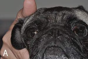
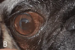
In a patient with severe lagophthalmos and resultant ocular disease, a procedure known as medial canthoplasty can help considerably (Figure 2).1-8 Although it does not correct all anatomic and physiologic features that threaten globe health and security, it can be very effective at reducing exposure and frictional irritation that can lead to keratitis and pigmentation. This procedure corrects the characteristics of brachycephalic ocular syndrome by:
- Reducing the length of both the upper and lower eyelids and overall length of the palpebral fissure
- Everting the medial inferior entropion
- Relieving functional obstruction of thenasolacrimal apparatus
- Removing the hairy medial caruncle and any additional medially deviated trichiasis. By improving globe coverage, effectiveness of the blink mechanism may be enhanced, tear fluid may be better distributed across the ocular surface, and the risk of proptosis may be reduced.
- Patients should wear an Elizabethan collar to prevent self- trauma and premature removal of sutures. If the incision site and eyelid structure break down, eyelid function and the globe will be compromised.
- Most patients benefit from topical broad-spectrum antibiotic therapy and a few days of systemic anti-inflammatory and, possibly, analgesic medications (Table).
- Sutures should be left in place for a full 14 days, earlier removal may cause incision failure.
More information can be found in this article